Research and development item: ③(c) Development of cell culture models of lung alveoli as evaluation systems for mathematical modeling
Implemented by the University of Tokyo
Final objective: We develop an in vitro alveolar model evaluation system using human cell lines and a primary rat culture system, establish a method to obtain various physicochemical and biological parameters for a mathematical model from its results, and propose an appropriate methodology.
In order to refine a mathematical model to describe and predict the intake, behavior, and impact of nanoparticles, we aim to establish an experimental model consisting of cultured cells for the alveoli, an intake route to the individual, and describe lesions and permeability with a simple mathematical model.
Main results:
With respect to the alveoli model using human cell line, on a commercially available semi-permeable membrane incubator (culture insert), we established a co-culture system of A549 cells with THP-1 cells derived macrophage-like cells. On a single A549 cell monolayer as an alveolar epithelial layer, THP-1 cells derived macrophage-like cells, as alveolar macrophage, were attached. We first confirmed that upon exposure to nanoparticles in the co-culture system, macrophage engulfed nanoparticles, resulting in less number of particles that are directly exposed on the epithelium, and lesions decreased. Regarding the permeability of nanoparticles, we conducted an experiment on the migration of nanoparticles, exposed to the inside of the alveoli (upper side of the co-cultured epithelium) over time. A temporal change in the quantity of nanoparticles in three compartments, i.e., inside the alveoli, inside the cell layer, and at the blood side (lower side of the co-cultured layer) in this experiment system, could be roughly explained with a mathematical model with dynamic equilibrium assumed among the three compartments. The permeability of the nanoparticles after 24 h was several percent or less; the permeability decreased significantly because of the presence of THP-1 cells derived macrophage-like cells.
As an in vitro evaluation system that functions in parallel with animal individual testing, we have been working on the construct and use of a primary rat culture in vitro alveolar model (Figure ③(c)). In the case of a primary rat culture, by seeding the epithelial type II cells, which could be purified, at an appropriate density on the culture insert, we successfully created an extremely thin continuous monolayer of the epithelium of type I cells that is similar to the actual inside of the alveoli. This monolayer exhibited a temporal change in its transepithelial electrical resistance value and the final cell shape. In the case of A549 cell line (II type), the area of individual cells was small, forming a dense monolayer; however, the electrical resistance value remained very low. However, primary culture alveolar epithelial cells changed approximately one week later to a shape specific to type I having a very large area. These cells created thin continuous monolayers similar to the alveoli; however, unlike the A549 type II-like cells, the electrical resistance value was very high, approximately ten times that of A549, and remained high for approximately one week. Thus, we created a very thin culture alveolar epithelium with a high barrier function similar to the actual in vivo alveolar epithelium. When we seeded alveolar macrophage collected separately on this epithelium, it attached firmly and remained stable, which demonstrates that we successfully established a primary co-culture the in vitro alveolar model. We are currently conducting experiments related to lesions and permeability of various nanoparticles and are proceeding with research to describe and evaluate the results using the mathematical model.
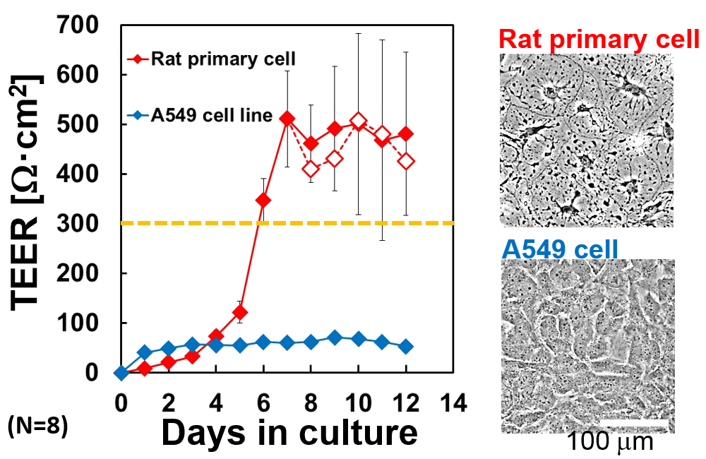
Figure③(c) Construction of primary rat culture in vitro alveolar model